Chromosome Analysis, Karyotyping

Our Chromosome Analysis service provides detailed insights into the structure and number of chromosomes in your samples. Trust us to deliver accurate results quickly, so you can focus on what matters most—your research.
Accepted cell types include:
- iPSC
- ESC
- Fibroblast
- Animal cells
- Cell fusions
And may be submitted as:
- Live cultures (adherent or suspension)
- Cryovials
- Harvested cell pellets
- Digital images
Purpose
Verification of a normal genome at the chromosomal level. G-banded karyotyping remains the ‘gold standard’ for examining the chromosomal structure of individual cells. This test detects aneuploidies and structural aberrations larger than 5Mb in size, such as: balanced translocations, inversions, deletions and duplications.
When to order
After multiple passages, before and after a manipulation, upon observation of unusual growth properties.
Methodology
Metaphase cell preparations are G-banded with trypsin and Wright’s stain. The chromosomes of individual cells are imaged and analyzed for aberrations.
Study parameters
ACMG (American College of Medical Genetics and Genomics) clinical analysis guidelines for neoplastic diagnoses, focused on the potential presence of low frequency/emerging mosaicisms within cell lines.
- 20 fully analyzed cells. Number of cells examined can be increased for higher sensitivity, upon request.
- When a non-clonal abnormality is observed, 20 additional cells are screened. Any cells identified with the same abnormality are fully analyzed and the abnormality is recategorized as ‘clonal’ and of genuine concern to the stability of the line.
Report
Comprehensive report provided using current ISCN nomenclature. Representative karyogram image of each clonal cell population identified.
Turn around time
Typically between 1-7 days. Dependent upon whether cultures need additional incubation time after delivery. Rush services available.
Fluorescence In Situ Hybridization (FISH)

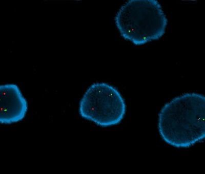
Our FISH service uses targeted probes to detect the presence and location of specific DNA sequences in individual cell nuclei.
Custom FISH studies
Please contact us to confirm probe availability prior to placing an order.
Purpose
In situ detection of specific loci. Probe sets are designed to identify specific abnormalities, such as: deletions, amplifications, cryptic rearrangements, and gene fusions.
When to order
The targeted nature of FISH makes it highly complementary to karyotyping and can identify aberrations that are otherwise cryptic. FISH is most often used as an adjunct to karyotyping, as the harvested material can be utilized for both tests. FISH may also be performed as a stand-alone test. Accurate quantification of mosaic cell populations is another strength of this method.
Methodology
Fluorescently labeled probes are hybridized to individual interphase or metaphase cells and the signal patterns are categorized.
Study parameters
Interphase FISH studies include a minimum of 200 cells and are particularly useful for the identification of amplifications, such as those involving BCL2L1 (20q). Metaphase FISH is typically used for confirmation, or more specific characterization of an abnormality identified by chromosome analysis.
Report
Comprehensive report provided using current ISCN nomenclature. Representative FISH image of each clonal cell population identified.
Turn around time
Rapid turnaround time, as short as 24 hours. Dependent upon the availability of harvested material and applicable probes.
Multi-Species Karyotyping

While not as commonly utilized as human iPSCs and ESCs, non-human and fusion cell lines remain of interest to researchers studying the unique cellular properties of various organisms.
GeneCode has extensive experience working with a wide range of species.
Copyright © 2020-2025 GeneCode Healthcare - CLIA ID# 05D2297708 - All Rights Reserved.
This website uses cookies.
We use cookies to analyze website traffic and optimize your website experience. By accepting our use of cookies, your data will be aggregated with all other user data.
